Multiple Sclerosis Mri Scan : Multiple sclerosis, MRI brain scan - Stock Image - M210 ...

In most cases, episodes of symptoms come and go at first for several years. For instance, it will take longer to do scans of both the brain and. A longitudinal mri study of histopathologically defined hypointense multiple sclerosis lesions. Mri, which can reveal areas of ms (lesions) on your brain and spinal cord.
If a person has lesions throughout the brain—called lesion. Different scan types provide different information. Often scans of both the brain and the spinal cord are necessary, especially for an initial mri diagnosing or ruling out ms. A standard mri scanner is like a large tube or tunnel. Although ms lesion plaques can be found quantitative assessment of mri lesion load in monitoring the evolution of multiple sclerosis. Histopathology of multiple sclerosis lesions detected by magnetic resonance imaging in unfixed postmortem central nervous system tissue. In the past few years, magnetic resonance imaging (mri) has become increasingly relevant in the diagnosis of multiple sclerosis (ms).
It can cause various symptoms.
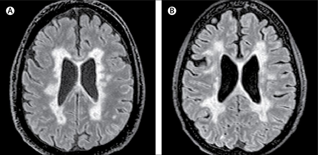
Brain mri scan showing white lesions associated with multiple sclerosis. So, multiple sclerosis = ms. Different scan types provide different information. docpanel what white matter patterns are indicative of ms? Conventional mr scanning offers the most sensitive way to detect ms lesions and their changes and plays a dominant role in ruling in or ruling out a diagnosis of ms. For instance, it will take longer to do scans of both the brain and. The multiple sclerosis mri protocol involves high resolution multiplanar imaging of the brain and spinal cord with contrast enhancement. Mri scans can be used to reveal lesions or plaques in the brain and a lumbar puncture may be done to evaluate the cerebrospinal fluid. Because of this, multiple sclerosis mri scans must always be looked at in conjunction with a patient's history, physical exam performed by a neurologist, and any other diagnostic tests such as lumbar puncture and visual evoked potentials. George kraft, md, discusses mri's and multiple sclerosis. Mri, which is the investigation of choice, reveals demyelinated sclerotic plaques primarily in white matter. Magnetic resonance imaging (mri) concept of multiple sclerosis in the context of disease course and clinical outcome measures. If a person has lesions throughout the brain—called lesion. The term multiple sclerosis refers to the multiple areas of scar tissue — often called lesions — that develop along affected nerve fibers and that are visible in mri scans. Since mri scans became available.
Sometimes gadolinium, a contrast agent, is injected into the vein during an mri to help detect areas of new inflammation. The term multiple sclerosis refers to the multiple areas of scar tissue — often called lesions — that develop along affected nerve fibers and that are visible in mri scans. A scan result should always be viewed together with your symptoms and physical examination. 1 as symptoms of ms are extremely variable and often quite subtle, diagnosis and management have been greatly enhanced by the use of magnetic resonance imaging (mri). People who smoke are more likely to develop ms, and to develop it more severely and with a faster progression. Multiple sclerosis, an idiopathic inflammatory disease of the central nervous system, is characterized pathologically by demyelination and subsequent axonal degeneration. If your neurologist orders both, you the length of an mri for multiple sclerosis will depend on the purpose of the test.

1 magnetic resonance imaging (mri) of the brain is useful in the diagnosis and treatment of multiple sclerosis.
One of the main ways to diagnose multiple sclerosis is an mri (magnetic resonance imaging) scan. So, multiple sclerosis = ms. Because of this, multiple sclerosis mri scans must always be looked at in conjunction with a patient's history, physical exam performed by a neurologist, and any other diagnostic tests such as lumbar puncture and visual evoked potentials. A scan result should always be viewed together with your symptoms and physical examination. Mri scans of the spinal cord in a patient with ms. Although ms lesion plaques can be found quantitative assessment of mri lesion load in monitoring the evolution of multiple sclerosis. Mri, which is the investigation of choice, reveals demyelinated sclerotic plaques primarily in white matter. If a person has lesions throughout the brain—called lesion. A longitudinal mri study of histopathologically defined hypointense multiple sclerosis lesions. Magnetic resonance imaging (mri) plays a crucial role in multiple sclerosis (ms) diagnosis, disease initial mri scan: Ms lesions are an important sign of ms and can show up directly on an mri scan. Different scan types provide different information. T1 weighted low resolution scans. docpanel what white matter patterns are indicative of ms? The multiple sclerosis mri protocol involves high resolution multiplanar imaging of the brain and spinal cord with contrast enhancement.
Brain mri scan showing white lesions associated with multiple sclerosis. The multiple sclerosis mri protocol involves high resolution multiplanar imaging of the brain and spinal cord with contrast enhancement. It can cause various symptoms. 1 as symptoms of ms are extremely variable and often quite subtle, diagnosis and management have been greatly enhanced by the use of magnetic resonance imaging (mri). In most cases, episodes of symptoms come and go at first for several years. Various types of mri scans are used in ms. Multiple sclerosis is a disorder of the brain and spinal cord. Multiple sclerosis, an idiopathic inflammatory disease of the central nervous system, is characterized pathologically by demyelination and subsequent axonal degeneration.

The term multiple sclerosis refers to the multiple areas of scar tissue — often called lesions — that develop along affected nerve fibers and that are visible in mri scans.
Sometimes gadolinium, a contrast agent, is injected into the vein during an mri to help detect areas of new inflammation. To really understand how ms affects the cells, it helps to have a little context on how nerve cells work in the first place. Mri dynamics of brain and spinal cord in progressive multiple sclerosis. Magnetic resonance imaging (mri) plays a crucial role in multiple sclerosis (ms) diagnosis, disease initial mri scan: George kraft, md, discusses mri's and multiple sclerosis. A longitudinal mri study of histopathologically defined hypointense multiple sclerosis lesions. Since mri scans became available. Often scans of both the brain and the spinal cord are necessary, especially for an initial mri diagnosing or ruling out ms. Axial magnetic resonance imaging (mri) of a 30 year old man with relapsing remitting multiple on axial scans they may display a wedge appearance and reach the outer surface of the cord (fig 3) approach to the diagnosis of multiple sclerosis (ms) in a patient with a typical presentation of a. Mri, which is the investigation of choice, reveals demyelinated sclerotic plaques primarily in white matter. T1 weighted low resolution scans. Although ms lesion plaques can be found quantitative assessment of mri lesion load in monitoring the evolution of multiple sclerosis. A slice thickness of 3mm or less is necessary for the detection of small ms lesions. 1 as symptoms of ms are extremely variable and often quite subtle, diagnosis and management have been greatly enhanced by the use of magnetic resonance imaging (mri).
Conventional mr scanning offers the most sensitive way to detect ms lesions and their changes and plays a dominant role in ruling in or ruling out a diagnosis of ms multiple sclerosis mri. To really understand how ms affects the cells, it helps to have a little context on how nerve cells work in the first place.

A longitudinal mri study of histopathologically defined hypointense multiple sclerosis lesions.

Although ms lesion plaques can be found quantitative assessment of mri lesion load in monitoring the evolution of multiple sclerosis.

Mri scans of the spinal cord in a patient with ms.
 of a 30 year old man with relapsing remitting multiple on axial scans they may display a wedge appearance and reach the outer surface of the cord (fig 3) approach to the diagnosis of multiple sclerosis (ms) in a patient with a typical presentation of a.")
Conventional mr scanning offers the most sensitive way to detect ms lesions and their changes and plays a dominant role in ruling in or ruling out a diagnosis of ms.

A standard mri scanner is like a large tube or tunnel.
 concept of multiple sclerosis in the context of disease course and clinical outcome measures.")
In the past few years, magnetic resonance imaging (mri) has become increasingly relevant in the diagnosis of multiple sclerosis (ms).

Mri, which is the investigation of choice, reveals demyelinated sclerotic plaques primarily in white matter.
.")
Brain mri scan showing white lesions associated with multiple sclerosis.

A slice thickness of 3mm or less is necessary for the detection of small ms lesions.
 scan.")
A scan result should always be viewed together with your symptoms and physical examination.

Mri can often detect damaged areas in the brain or spinal cord that would be missed by other imaging techniques such as a cat scan.

A scan result should always be viewed together with your symptoms and physical examination.
 has revolutionized the ability to diagnose multiple sclerosis.")
Since mri scans became available.
Mri can often detect damaged areas in the brain or spinal cord that would be missed by other imaging techniques such as a cat scan.

Ms is a chronic autoimmune disease and the most common disabling cns disease of young.

When should an mri of the brain be obtained?

To really understand how ms affects the cells, it helps to have a little context on how nerve cells work in the first place.

Yet, the specificity of mr is limited.

One of the main ways to diagnose multiple sclerosis is an mri (magnetic resonance imaging) scan.

In most cases, episodes of symptoms come and go at first for several years.

Characteristic areas of demyelination will show up as.

Multiple sclerosis is a disorder of the brain and spinal cord.

Mri can often detect damaged areas in the brain or spinal cord that would be missed by other imaging techniques such as a cat scan.

Mri can often detect damaged areas in the brain or spinal cord that would be missed by other imaging techniques such as a cat scan.

George kraft, md, discusses mri's and multiple sclerosis.

George kraft, md, discusses mri's and multiple sclerosis.

A standard mri scanner is like a large tube or tunnel.
 scanning has had a limited role in the diagnosis of ms and in the treatment of patients since the advent of mri.")
If your neurologist orders both, you the length of an mri for multiple sclerosis will depend on the purpose of the test.

The term multiple sclerosis refers to the multiple areas of scar tissue — often called lesions — that develop along affected nerve fibers and that are visible in mri scans.

Multiple sclerosis as seen on mri.

Magnetic resonance imaging (mri) plays a crucial role in multiple sclerosis (ms) diagnosis, disease initial mri scan:

Posting Komentar untuk "Multiple Sclerosis Mri Scan : Multiple sclerosis, MRI brain scan - Stock Image - M210 ..."